 |
| katsumi�@Saitoh*1�@Hajime Muto�C2�@ Noriyuki Hachiya�C3 and�@Yukio Takizawa3 |
| 1Division of Environmental Science�C Akita Prrefectural Institure for Fisheries
and Fisheries Management,Uosaki 16,Dashima,Funagawahou,Oga-shi,Akita 010-05,Japan; 2Environmental Research Center,Akita University,Akita 010, Japan,and 3Department of Public Health,Akita University School of Medicine, Akita 010,Japan |
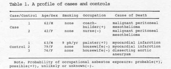
It has longbeen�@known�@that�@diffuse�@interstitial�@pulmonary�@fiberrosis can be caused by asbests(Doll and Petro 1985). Definite exposure reponse relations between both level and duration of exposure to asbestos and presence of difinite radiographic abnormalities have been shown by many authors (Finkelstein and Vinglis 1984; copes at al.1985).Asbestos bodies in lung tissue have been recognized as a marker of past asbestos exposure(Churg and Warnoc 1977). Although there is no convincing evidence that indirect exposure contributes to the cccurrence of mesothelinomas, there have been numerous reports of this rate tumor in dividuals exposed to asbestos(Anderson et al.1976;Vianna and Polan 1978). From the review of all causes newly diagnosed in 1982 as a malignant mesothelioma of the pleura or peritoneum(Churg 1985), it has been shown that the incidence rate of mesothelioma in British Columbia has increased nearly six times for men compared to the period 1969 to 1975,but remained roughly unchanged for women, and almost all of the case in men in this series could be linked to asbestos exposure. de Klerk et al.(1989)have predicted future incidence of asbestos-related disease in former Wittennoom asbestos workers in Western Australia for the period 1987 to 2020. They predicated 2893 deaths in this period, 692 cases of mesothelioma, 183 cases of lung cancer, and 482 cases of�@asbestosis. Additionally,they indicated that the incidence of both lung cancer and asbestosis was greatest in those subjects with the highest levels of exposure to crocidolite and in smokers(de Klerk et al.1991). Berry(1991)also predicted from the follw-up study on Wittenoom workers that between 250 and 500 deaths and between 340 and 465 deaths will occur due to mesothelioma and lung cancer,respectively. It is important for mesothelioma to have criteria other than histry by which a case may be classified as asbestos-related(Warnoc 1989). The risk of malignant mesothelioma associated with low-level asbestos exposure is an important unresolved issue tody(Mowe et al.1985). We report here a case study on malignant peritoneal mesothelioma associated with asbestos, using a scanning electron microscope with x-ray microanalyzer and a phase-contrast microscope. MATERIALS AND METHODS The source population for both cases and controls was the population of Akita city in the North part of Japan in 1987. The incidence rate per year of carcinomatous perritonitis including malignant peritoneal mesothelioma in Japan has been reported as about 3.3per million for general population from 1985 to 1989(Ministry of Health and Welfare,Japan 1985-1989). Two patients diagnosed as malignant peritoneal mesothelioma in Akita Kumiai General Hospital were subjected, and three populations who died of mycardial infarction or dissecting aortic aneurysm in the hospital were chosen as controls. Table1 shows their profiles. Analytical procedure used to determine asbestos bodies and fibers in pathological autopsy samples has been described elsewhere(Ashcroft and Heppleston 1973). About 5g of the sample was homogenized with distilled water at high speed in a Ultrahomogenizer(Physcotron:Ikemoto Sci. Technol.Co.,Japan). An aliquot of the sample was transferred to a centrifugal tube of 50ml equipped with a condenser and saponified with 20ml of 40% KOH-ethanol solution in water bath at 100�� for an hour. After cooling, the sample was filtrated through a membrane filter(millipore,AA 0.8t��mx47 mm) with 150ml of distilled water. Asbestos sample trapped on filter was kept in a desicator for the determination using a phase-contrast microscope(Olympus,BH-I,Japan) and a scanning electron microscope (Hitachi s-7000,Japan)equipped with an energy-dispersive x-ray microanalyzer(Kevex,Delta�V,USA). Prior to the determination, asbestos sample was prepared by the acetone-triacetin method reported elesewhere (Japan Asbestos Assoc.1988). The dry weight of sample was equal to one-half of wet one. Chemicals were of reagent quality and abtained from Wako Pure Chemical Industries(Japan). |
 |
| Table2. Concentrations of asbestos bodies and naked fibers in lung, greater omentum, and large intestinal tissue samples from two patients with malignant peritoneal mesothelinoma and three general populations |
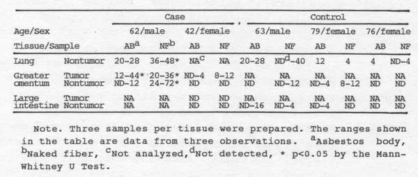 |
| RESULT AND DISCUSSION Asbestos body and fiber concentrations in lung, greater omentum, and large inteastine tissues of cases and controls are shown in Table2. For 62-years-old male of case 1, asbestos body concentrations in lung tissues ranged from 20 to 28 per g dry base and corresponded to the level which Zhang(1987)has classified to the slight exposure level by asbestos body as the ranges of 11 to 100per g wet lung. Asbestos body size of 100��m at the maximum was observed. The neked fiber concentrations in lung tissues were apporoximately two times, compared to those of asbestos bodies. For greater omentum, the bodies and fibers were also found in tumor or nontumor tissues. Their concentrations in tomor tissues were similar to the lung tissue levels, and their sizes were about 20��m for body and a few hundred ��m for fiber. |
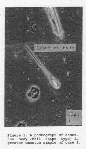 |
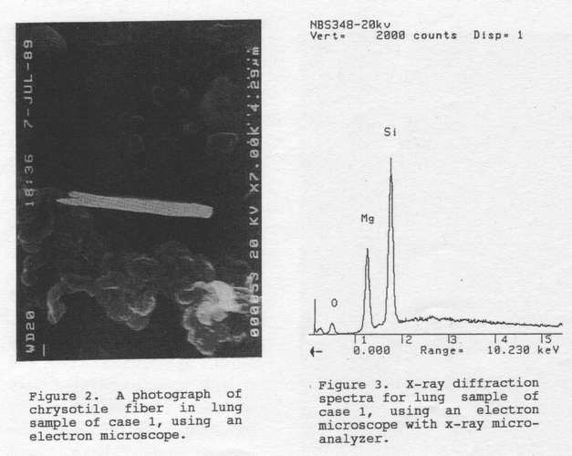
| Most of body types were bell and club shape types. A phase-contrast microscopic photograph of asbestos body in greater omentum tissue of case 1 is shown in Figure 1. Fiber concentrations for lung and greater omentum of case 1were significant(p<0.05> as compared with controls, using the Mann-Whitney U-tests. Furthermore, chrysotile fibers were identified in lung tissues of case 1, using the scanning electron microscope with energy-dispersive x-ray microanalyzer (see Figures 2 and 3). However, in order to the small number of patients and the lack of specimens of greater omentum and large intestinal tumor samples in the control patients, it is not clear whether or not the differences in fiber concentrations between case 1 and controls sre shown. |
 |
|
Fiber size is believed to play a role in determining risk of a particular asbestos-related disease (Lippmann 1988). Warnock(1989) has quantified the lung asbestos burder in shipyard and construction workers with mesothelinoma, and reported that their burder was significantly greater than the burder found in men of general populations(p<0.01). Furthermore, because that the median concentration for total amphibole fibers in subjects with mesothelinoma did not differ significantlly as compared with subjects with asbestosis, it was hypothesized that fiber size, especially amosite of the most prevalent type, would differ among asbestos-related disease. For our case study, it was suggested that the significant difference in fiber concentrations in greater omentum tissues of a patient(case 1) with malignant peritoneal mesothelioma would associate with asbestos exposure . Then, the pleural plaques were not observed from pathological findings of case 1. However,for the relation between asbestos fiber types and pleural plaques in a general autopsy population, it has been suggested that the presence of pleural plaques correlates with a modest(50-fold) increase in numbers of long high-aspect ratio commercial amphiboles(amosite and crocidelite) in lung tissue(Churg 1982). |
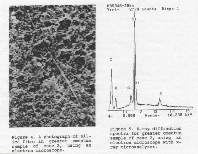 |
For 42year-old female of case 2, although a few numbers of asbestos bodies and fibers were detected in tumor tissues of greater omentum, their concentrations were much low, and the body types were similar to case 1, Howwver , they were not observed in nontumor tissues of greater omentum and in tumor and nontumor tissues of large intestine. On the other hand , a large quantity of fiberous substances of needle-type were observed in greater omentum tissues. It was found that the materials were silica fibers (see Figures 4 and 5), using the scanning electron microscope. Silica fibers were not in above tissues of case 1 and controles. Considering that mesothelioma is also induced by the stable and fiberous substances other than asbestos in vivo (Stanton et al.1981), it was suggested that asbestos as well as fiberous substances such as silica fibers might play an important role malignant peritoneal mesothelioma. REFERENCES Anderson HA,Lilis R,Daum SM, Fischbein AS, Selikoff IJ(1976) Household-contact asbestos neoplastic risk. Ann NY Acad Sci 271:311-23 Ashcroft T, Heppleston AG(1973) The optical and electron microscopic determination of pulmonary asbestos fiber concentration and uts relation to the human pathological reaction. J Clin Pathol 26: 224-34 Berry G(1991) Prediction of mesothelioma, lung cancer, and asbestosis in former Wittennoom asbestos workers. Br J Ind Med 48: 793-892 Chrg A, Warnock ML(1977) Correlation of quantitative asbestos body counts and occupation in urban patients.Arch Pathol Lab Med 101:629-34 Chrg A,(1982) Asbestos fibers and pleural plaques in a general autopsy population. Am J Patol 109: 88-96 Chrg A,(1985) Malignant mesothelioma in British Columbia in 1982. Cancer 55:672-4 Copes R, Thomas D, Becklate MR(1985) Temporal patterns of exposure and nonmalignant pulmonary abnormality in Quebec chrysotile workers. Arch Environ Hlth 40:80-7 de Klerk NH, Armstrong BK, Musk AW, Hobbs MST(1989) Prediction of future cases of asbestos-related disease among former miners and millers of crocidolite in Western Australia. Med J Aust 151:616-20 de Klerk NH, Musk AW, Armstrong BK, Hobbs MS(1991) Smoking,exporsure to crocidolite, and the incidence of lung cancer and asbestosis. Br J Ind Med 48:412-7 Doll R, Petro J(1985) Asbests:Effects on health of exposure to asbestos.HMSO,London Finkelstein MM,Vingilis JJ (1984) Radiographic abnormalities among asbestos-cement workers.Amer Rev Respir Dis 129:17-22 Japan Asbestos Association(1988) Analytical method of asbestos concentration in room air.Japan Association for working Environmental Measurement,4:6-12,Tokyo Lippmann M(1988) Asbestos exposure indices. Environ Res 46:86-106 Ministry of Health and Welfare, Japan(1985-9) Vital statistics,Department of Statistics and Information of Ministers Secretariat,Tokyo Mowe G, Gylseth B,Hartveit F Skaug V (1985) Fiber concentration in lung tissue of patients with malignant mesothelioma. Cancer 56:1089-93 Stanton MS,Layard M,Tegeris A,Miller E,May M, Morgan E, Smith A(1981) Relation of particle dimension to carcinogenicity in amphibole asbestoses and other fiberous minerals. J Natl Cancer Inst 67:965-75 Vianna NJ,Polan AK(1978) Nom-occupational exposure to asbestos and malignant mesothelioma in females.Lancet:1061-3 Warnock ML(19899 Lung asbestos burden in shipyard and construction workers with mesothelioma :Comparison with burdens in subjects with asbestosis or lung cancer. Environ Res 50:68-85 Zhang W(1987) A quantitive study on asbestos exposure to the extra pulmonary organs:With special reference to the extra pulmonary effect of asbestos. The Niigata Med J Jpn 101:116-25 Received May 6 1992; accepted September 2 1992. �A�X�x�X�g���g�b�v�y�[�W�� ���S�Ȋw�A�J�f�~�[�g�b�v�y�[�W�� |